Epizootic Hemorrhagic Disease
Other Names: Hemorrhagic disease
Cause
Hemorrhagic Disease (HD) in white-tailed deer can be caused by either of two closely related viruses, Epizootic Hemorrhagic Disease Virus (EHD) and Bluetongue Virus (BT). Outbreaks of diseases similar to HD have been described since 1890, but the EHD virus that causes this disease was not isolated until an outbreak in New Jersey white-tailed deer in 1955. The BT virus was isolated from white-tailed deer and bighorn sheep with HD in Texas in 1966. So far HD in Pennsylvania has been caused by the EHD virus.
Significance
Hemorrhagic disease is one of the most common diseases of white-tailed deer in the eastern United States and can cause a significant number of deaths during outbreaks.
Species Affected
White-tailed deer and mule deer are the primary wildlife species affected by HD. This disease is occasionally associated with deaths of pronghorn antelope and bighorn sheep. Elk can become infected with this disease, but they do not seem to be nearly as susceptible as white-tailed deer. EHD rarely causes disease in domestic animals, while BT is a well-known disease of sheep, cattle, and goats and can also infect domestic dogs. The viruses are not known to cause disease in humans.
Distribution
EHD and BT viruses are found worldwide in temperate and tropical climates, but they have only been reported in free-ranging wildlife in North America. In the United States, HD has been confirmed in most eastern and southeastern states as well as several states in the Midwest and northwest. There have also been sporadic cases reported in British Columbia, Alberta, and Saskatchewan in Canada. Pennsylvania has recently had three outbreaks of hemorrhagic disease in white-tailed deer in 1996 (not confirmed), 2002, and 2007. One positive case was diagnosed in Northampton county in August of 2011.
Transmission
The EHD and BT viruses are both transmitted by biting flies or midges in the group called Culicoides (Ku/lick/oyed/eez). Female midges pick up the viruses by ingesting the blood of an infected animal and they then transmit the viruses when they feed on an uninfected animal. Midges are found near mud, which is their preferred breeding habitat, so outbreaks usually occur when deer congregate in wet areas at the driest part of late summer and early fall when seasonal midge activity is also at its peak. They end when the first hard frosts take their toll on the midges and the virus dies with the insects.
The species of Cullicoides that is considered the primary vector of EHD is not thought to normally occur in Pennsylvania, but is occasionally brought into the state on wind currents. As a result, Pennsylvania deer do not have any immunity to the virus because outbreaks usually occur at intervals which are longer than the average deer's lifespan. This lack of immunity explains the high mortality rates during EHD outbreaks in Pennsylvania.
Clinical Signs
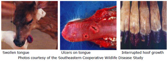
Clinical signs of hemorrhagic disease are all a result of the damage that the virus does to the walls of the blood vessels. They can range from sudden death to chronic disease. White-tailed deer usually develop clinical signs of HD about 7 days following infection with the EHD or BT virus, but some animals may remain asymptomatic. Clinical signs include swelling of the face or neck, loss of appetite, lethargy, weakness, lameness, respiratory distress, fever, and excessive salivation. Deer with HD will often have ulcers in the mouth and may bleed from the nose and/or mouth. Infected animals may develop swollen, blue tongues. They will also often experience hoof overgrowth and may have indentations or cracks in the walls of their hooves (see photos). Usually infected deer will go into shock and die within 8 to 36 hours of the onset of clinical signs. Necropsy of animals that die of HD will often reveal extensive hemorrhage from any or all internal organs. Organs more likely to exhibit hemorrhage include the heart, liver, kidneys, lungs, spleen, and intestines.
Diagnosis
Laboratory tests are used to isolate the EHD or BT virus from infected tissues. Blood, liver, spleen, kidney, lung, heart, and muscle tissue can all be used for virus isolation.
Treatment
There is currently no treatment for hemorrhagic disease in wildlife populations.
Management/Prevention
Hemorrhagic disease can cause very high mortality rates and is considered the most important viral disease of white-tailed deer in the United States. Both wild free-ranging and captive deer and elk are at risk of contracting HD, and the disease can be spread by transporting infected animals to areas where the disease is not yet present. The potential impact of this disease on deer populations is currently unknown, but it is important to test suspect cases in order to identify outbreaks of HD. Insect control could potentially decrease transmission of EHD and BT viruses in captive herds, but it is not feasible in wild populations.
Suggested Reading
Bluetongue. General Disease Information Sheets. OIE: World Organization for Animal Health. http://www.oie.int/en/animal-health-in-the-world/disease-information-summaries.
Howerth, E. W., D. E. Stallknecht, and P. D. Kirkland. 2001. Bluetongue, epizootic hemorrhagic disease, and other orbivirus-related diseases. Pages 77-97 in E. S. Williams and I. K. Barker, editors. Infectious diseases of wild mammals. Iowa State University Press, Ames, Iowa, USA.
Michigan Department of Natural Resources. Wildlife Disease. Epizootic hemorrhagic disease (EHD) in white-tailed deer. http://www.michigan.gov/dnr/0,1607,7-153-10370_12150_12220-26647--,00.html.
Sleeman, J. M., J. E. Howell, W. M. Knox, and P. J. Stenger. 2009. Incidence of hemorrhagic disease in white-tailed deer is associated with winter and summer climatic conditions. EcoHealth 6: 11-15.